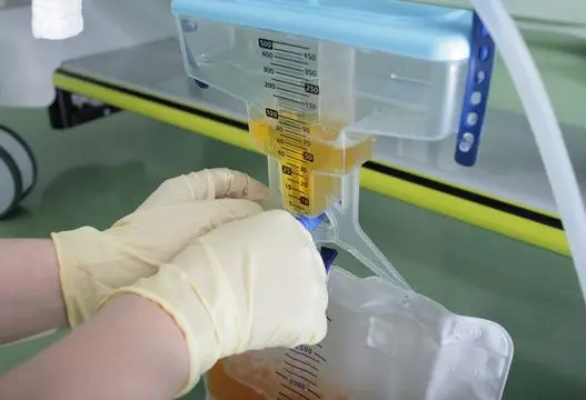


Drug development timelines require reliable data for IND or NDA submissions, making the selection of an immunoassay critical. Two common methods for pharmacokinetic testing are traditional enzyme-linked immunosorbent assays (ELISA) and Meso Scale Discovery (MSD) platforms. While ELISA has been the standard, newer electrochemiluminescence (ECL) technologies offer enhanced performance. This guide compares the operational differences between MSD ECL assays and traditional ELISA, focusing on sensitivity, dynamic range, and data reliability for GLP-compliant bioanalysis in complex biological matrices.
Defining the MSD ECL Assay and Traditional ELISA
Standard enzyme-linked immunosorbent assays rely on colorimetric or fluorescent signals to measure analyte concentrations. A capture antibody binds the target, and an enzyme-linked detection antibody generates a signal proportional to the analyte concentration. While reliable for many applications, this method often requires large sample volumes and multiple dilution steps.
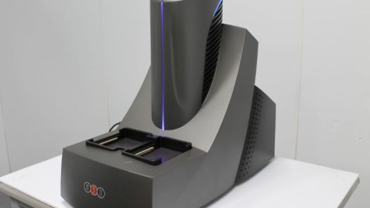
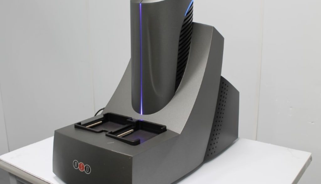
The MSD ELISA assay alternative utilizes an electrochemiluminescence (ECL) detection system. Instead of an enzyme, the detection antibody is labeled with ruthenium. When an electric field is applied to the microplate electrodes, the ruthenium label emits light. This reaction separates the excitation mechanism from the emission signal, which drastically reduces background noise. 
Direct Comparison of Sensitivity Levels
Sensitivity is a primary concern during assay validation, especially when measuring low-abundance biomarkers or therapeutics. The detection limits of your chosen method dictate the reliability of your Pharmacokinetic Testing data.
- Standard Platforms: Traditional colorimetric assays typically detect analytes at picogram-per-milliliter (pg/mL) concentrations. This level of sensitivity works well for many standard bioanalysis projects.
- ECL Platforms: An MSD immunoassay routinely achieves sub-picogram-per-milliliter sensitivity. The electrical stimulation of the ruthenium label produces a high-intensity signal with minimal background interference.
This higher sensitivity allows researchers to accurately quantify targets that fall below the lower limit of quantification (LLOQ) of older methods. For teams developing novel biologics, this capability provides clearer safety and efficacy data.
Analyzing Dynamic Range and Mesoscale Cytokine Multiplex Technology
A broad dynamic range reduces the need for multiple sample dilutions, saving time and conserving limited sample volumes.
- Traditional Assays: Offer a dynamic range of two to three logs, often requiring serial dilutions for high-concentration samples. This process increases manual labor and the risk of technical variability.
- MSD Assays: Provide a dynamic range of three to five logs. This allows measurement of both high- and low-concentration samples on the same plate, minimizing the need for dilutions.
The platform also supports efficient multiplexing using mesoscale cytokine multiplex technology. This enables researchers to measure up to ten different analytes in a single well with only 25 microliters of sample. This is particularly beneficial for pediatric studies or rare-disease trials where sample sizes are limited.
Benefits of the MSD Cell-Based Assay in Drug Development
Modern therapeutics often require biologically relevant cellular testing to confirm target engagement and potency. Researchers frequently use cell-based assays to evaluate mechanisms of action and neutralizing antibody responses.
Implementing an MSD Cell-based Assay offers several distinct operational benefits:
- Reduced wash steps: The ECL format involves fewer wash cycles than standard protocols, thereby preserving delicate cell monolayers.
- Direct measurement: Scientists can measure intracellular signaling proteins directly in the assay plate, without extraction.
- High throughput: The platform supports rapid plate reading, which accelerates data acquisition for large clinical cohorts.
These features streamline the workflow for drug development programs, ensuring that testing stays on schedule.
Technical Advantages in Reducing Matrix Effects
Biological matrices such as serum, plasma, and tissue lysates contain numerous endogenous compounds that can interfere with target detection. This phenomenon, known as the matrix effect, can significantly affect data accuracy. Conventional assays are particularly vulnerable, as colourimetric or fluorescent signals may be distorted by factors such as sample turbidity or inherent auto-fluorescence.
Electrochemiluminescence methods help reduce these challenges. The signal is generated through electrical stimulation, and the ruthenium label emits light at a specific wavelength, limiting interference from background autofluorescence. In addition, the carbon surface of the microplates enables strong protein binding, supporting thorough washing steps that remove non-specific matrix components before signal detection.
Must Read: Pharmacokinetics Assays for Small Molecules vs. Biologics
Conclusion
Both ELISA and MSD ECL assays play important roles in bioanalysis, but their performance differs significantly in terms of sensitivity, dynamic range, and resistance to matrix interference. While ELISA remains a reliable and widely used method, MSD ECL technology offers improved detection limits, a broader dynamic range, and efficient multiplexing capabilities. These advantages make MSD particularly suitable for complex biological matrices and studies involving low-abundance analytes. Selecting the appropriate platform ultimately depends on the study requirements, but advanced ECL systems can provide more precise, efficient data to support drug development and regulatory submissions.